The limbic system is comprised of both cortical and subcortical brain structures that play an important role in emotions, drives, olfaction, homeostasis, autonomic control, neuroendocrine control and memory. (I use the mnemonic “A Demon” or “Moaned” to remember these six functions)
Most of the limbic structures lie hidden in the ventral-medial areas of the brain and diagrams of them can be seen below. A list of the various structures of the limbic system can also be seen in the table to the right.
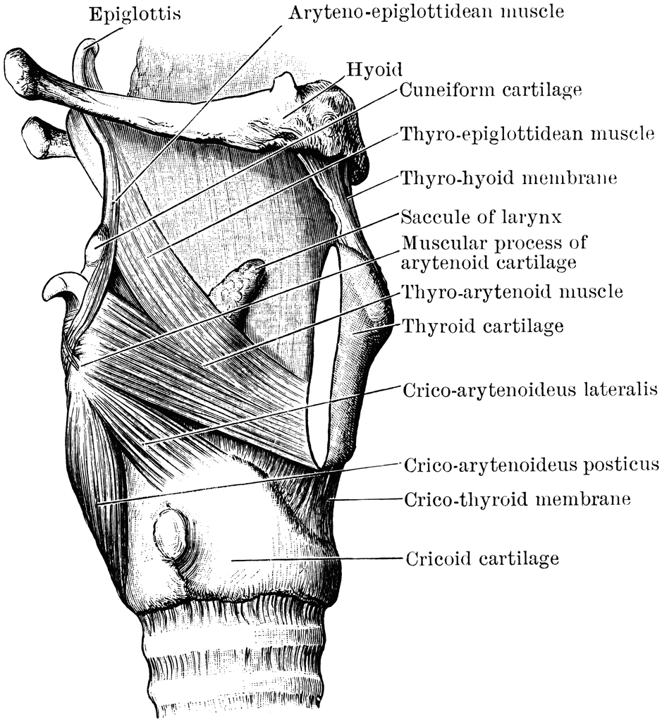
The olfactory system, or rhinencephalon, consists of several types of neurons. Olfactory receptors of the (1) olfactory nerves relay information about odorants into the olfactory bulb where they synapse with (2) mitral cells and (3) tufted cells. These cells relay information about the odorants through the olfactory tract and to the olfactory cortex. Additional neurons called (4) periglomerular cells and (5) granule cells participate in this process. Eventually information is received by the primary olfactory cortex, near the medial, anterior tip of the temporal lobes. The primary olfactory cortex is comprised of the piriform and periamygdaloid cortices. Information received by the primary olfactory cortex is then projected to several secondary olfactory areas such as the basolateral amygdala, mediodorsal nucleus of the thalamus, anterior entorhinal cortex and ultimately, the orbitofrontal olfactory areas.
The hippocampal formation is one of many different memory structures that make up the limbic system. It’s three components are the (1) dentate gyrus, (2) hippocampus and (3) subiculum. The anterior division of the hippocampus, known as the pes hippocampi, is the largest. As it progresses posteriorly it tapers and wraps posteriorly around the corpus collosum until it disappears under the splenium of the corpus callosum. A vestigial remnant continues along the dorsal surface of the corpus calossum, forming the indusium griseum.
The hippocampal formation is unique in that it is comprised of a special type of cortical tissue named archicortex. Archicortex is more phylogenetically ancient form of cortex that is comprised of only three layers (molecular, pyramidal and polymorphic layers in the hippocampal formation). The majority of our cerebral cortex is comprised of six layered neocortex.
A great deal of research has been compiled on how the circuitry of the hippocampal formation contributes to memory functions. Research suggests that pyramidal cells of the entorhinal cortex project to the hippocampal formation through the perforant and alvear pathways.
The parahippocampal gyrus includes several adjacent areas of cortex such as the entorhinal cortex (the major input/output relay between the association cortex and the hippocampal formation), the perirhinal cortex, piriform cortex, periamygdaloid cortex, presubicular cortex, prorhinal cortex and parahippocampal cortex. As the parahippocampal gyrus travels posteriorly it tapers into the parahippocampal cortex and then into the rhinal sulcus.
The fornix is a C-shaped collection of white matter tracts that serve as a major output pathway from the subiculum of the hippocampus to the septal nuclei and mammillary bodies. A smaller group of cholinergic neurons travel in the opposite direction from the medial septal nuclei and diagonal band of Broca, to the hippocampal formation.
The amygdala lies just dorsal to the anterior tip of the hippocampus and plays roles in emotion, homeostasis, memory and olfaction. However, it is best known for it’s prominent role in emotions and drives. The amygdala is comprised of (1) corticomedial nuclei (2) basolateral nuclei (3) central nuclei and (4) bed nuclei of the stria terminalis. The corticomedial nuclei are involved in olfaction and hypothalamic appetitive states. The central nuclei are also involved with the hypothalamus, in addition to the brainstem and play an important role in autonomic control. While activity in the amygdala is important in fear, anxiety and aggression, the septal area appears important for pleasureable states. The amygdala plays an important role in attaching emotional significance to memories. Finally, it also appears to have a role in neuroendocrinological functioning during states of altered emotional experience.
Diencephalic limbic structures (e.g. the hypothalamus, select thalamic nuclei and the nucleus accumbens) play a role in all limbic functions.
The septal nuclei play an important role in memory encoding. The lateral septal nuclei recieve information from from the precommissural fibers of the fornix. The medial septal nuclei send acetylcholine back in the opposite direction. This acetylcholine then allows memories to be formed by enabling theta electrical waves in the temporal regions.
Brainstem nuclei (i.e. interpeduncular nucleus, superior central nucleus, dorsal tegmental nucleus, ventral tegmental nucleus, parabrachial nucleus, periaquedctul gray, reticular formation, nucleus solitarius and the motor nucleus of the vagus) exert an influence on limbic pathways